The longevity-associated BPIFB4 gene supports cardiac function and vascularization in aging cardiomyopathy

Professor Paolo Madeddu and his team are working to delay the ageing of the heart. He tells Dr Leanne Grech how this research could allow older people to live a healthier life for longer.

The BPIFB4 gene has been associated with exceptional longevity

Each day, your heart beats around 100,000 times, pumping about eight pints of blood around your body. By the time you are 20, the heart’s function can begin to decline as part of normal ageing. As you get older, activities like running or playing tennis become more difficult. However, some 100-year-olds, like those living in Okinawa, a cluster of islands in southern Japan, appear to have unlocked the secret to a long and healthy life, with some of them seemingly having a heart younger than their age. “It’s a combination of a good lifestyle and good genes,” explains Professor Paolo Madeddu at the University of Bristol. “And we have discovered that one of these good genes can stop ageing.”

The gene that can stop ageing Professor Madeddu and his team have discovered that a naturally occurring variant of the BPIFB4 gene, which is more common in people who live to 100 or more, could help keep the heart young. A gene variant is a permanent change in the DNA sequence that makes up a gene. The team have already learned that mice treated with this variant have healthier hearts. In fact, transferring this gene variant to old mice seemed to help relax blood vessels in mice with high blood pressure and increase the amount of blood delivered to the muscles. In humans, ageing can affect many parts of the body, including weakening the heart and circulatory system. For example, heart failure, a serious and sometimes disabling condition for which there is no cure other than a heart transplant. Symptoms include breathlessness and feeling abnormally tired. Heart failure can occur at any age, but it is more common in older people, and in people who have had a heart attack and who have cardiomyopathy or high blood pressure.

Scientists are now beginning to understand how some natural variations in our genes might protect against heart diseases linked to ageing, such as heart failure. In this study, Professor Madeddu and his team in Bristol have been funded by the BHF with more than £172k to further investigate the role of the BPIFB4 gene variant. The BPIFB4 gene has been associated with exceptional longevity, helping protect against atherosclerosis (build-up of fatty material inside your arteries) and high blood pressure. The team now want to know if this gene variant can be given as a tablet and if it can reduce chronic inflammation (a damaging set of processes often seen in ageing hearts). Most genes contain the information needed to make functional molecules called proteins. Giving the corresponding protein as a pill instead of the gene variant could be an easier way to get the same result. Professor Madeddu and his team will test this idea in mice. If it succeeds, they hope to go on to a clinical trial in humans. “The study will provide proof of concept that our solution is valid. More studies are needed to show it is also safe – the fact that it is a human protein and not a [new] drug is encouraging. However, producing large quantities of protein is extremely expensive, and we will need investors or an industrial partner in the future. The BHF could help us to find the right partner,” explains Professor Madeddu.

I hope that the results of my work can make a difference in the lives of a lot of patients.

Helping older hearts Professor Madeddu is a cardiologist by background, and has been fascinated by the heart since he was a medical student. “Research requires a lot of determination and effort. I hope that the results of my work can make a difference in the lives of a lot of patients.” In 2021, around 12.5 million people in the UK were 65 or older. Life expectancy had been rising for decades, but the increases have slowed since 2010, and life expectancy for both men and women has fallen since the Covid-19 pandemic. Research like that of Professor Madeddu aims to help older adults stay healthy and independent for longer, which is among the UK Government priorities. “There is no current treatment to stop the heart’s ageing, and drugs used for heart disease can cause side effects in seniors,” explains Professor Madeddu. “We hope that giving older people a protein that is present in healthy centenarians, like those in Okinawa, helps their hearts work better for longer. Our approach proposes to increase health rather than simply combat disease. If we can find an effective treatment to delay ageing of the heart, we may be able to prevent serious disease in older people.”

Read the case report

Monica Cattaneo , Antonio P Beltrami , Anita C Thomas , Gaia Spinetti , Valeria Alvino , Elisa Avolio , Claudia Veneziano , Irene Giulia Rolle , Sandro Sponga , Elena Sangalli , Anna Maciag , Fabrizio Dal Piaz , Carmine Vecchione 4,5 , Aishah Alenezi , Stephen Paisey , Annibale A Puca , Paolo Madeddu The longevity-associated BPIFB4 gene supports cardiac function and vascularization in aging cardiomyopathy.  Cardiovasc Res. 2023 Jan 13;cvad008. PMID: 36635236 DOI: 10.1093/cvr/cvad008.

Stem cell plasters to stop children needing repeated heart surgeries

Researchers at the University of Bristol, funded by the British Heart Foundation (BHF), have developed ‘stem cell plasters’ to revolutionise the way surgeons treat children living with congenital heart disease, so they don’t need as many open-heart operations.

Heart defects are the most common type of anomaly that develop before a baby is born, with around 13 babies diagnosed with a congenital heart condition every day in the UK.  These include defects to the baby’s heart valves, the major blood vessels in and around the heart, and the development of holes in the heart.

Currently, for many of these children, surgeons can perform open-heart surgery to temporarily repair the problem, but the materials used for the patches or replacement heart valves are not completely biological and cannot grow with the baby. This means they can be rejected by the patient’s immune system which causes the surgical materials to gradually break down and fail in a few months or years.

A child might therefore have to go through the same heart operation multiple times throughout its childhood, which keeps them in hospital for weeks at a time, hugely impacts their quality of life and causes a lot of stress for the family.

Now, BHF Professor Massimo Caputo has developed the first type of stem cell patch to repair abnormalities to the valve in the large blood vessel that controls blood flow from the heart to the lungs, and to mend holes between the two main pumping chambers of the heart.

The stem cell plasters are designed to be sewn into the area of the child’s heart that needs repairing during surgery. The stem cells could then boost the repair of heart tissue without being rejected by the child’s body.

These patches have the potential to adapt and grow with the child’s heart as they get older, removing the need for repetitive heart surgeries and the many days at hospital recovering after each one.

There are around 200 repeat operations for people living with congenital heart disease every year in the UK. The technology could save the NHS an estimated £30,000 for every operation no longer needed, saving millions of pounds each year.

The BHF has awarded Professor Caputo nearly £750,000 with the aim to get these patches ready for testing in patients so clinical trials can start in the next two years, enabling more children and babies to benefit from the life-altering technology. The materials have already proven to work safely in animals.

The team is also in the early stages of developing other stem cell technologies using 3D bioprinting and gene therapy to one day be able to mend more complex congenital heart defects.

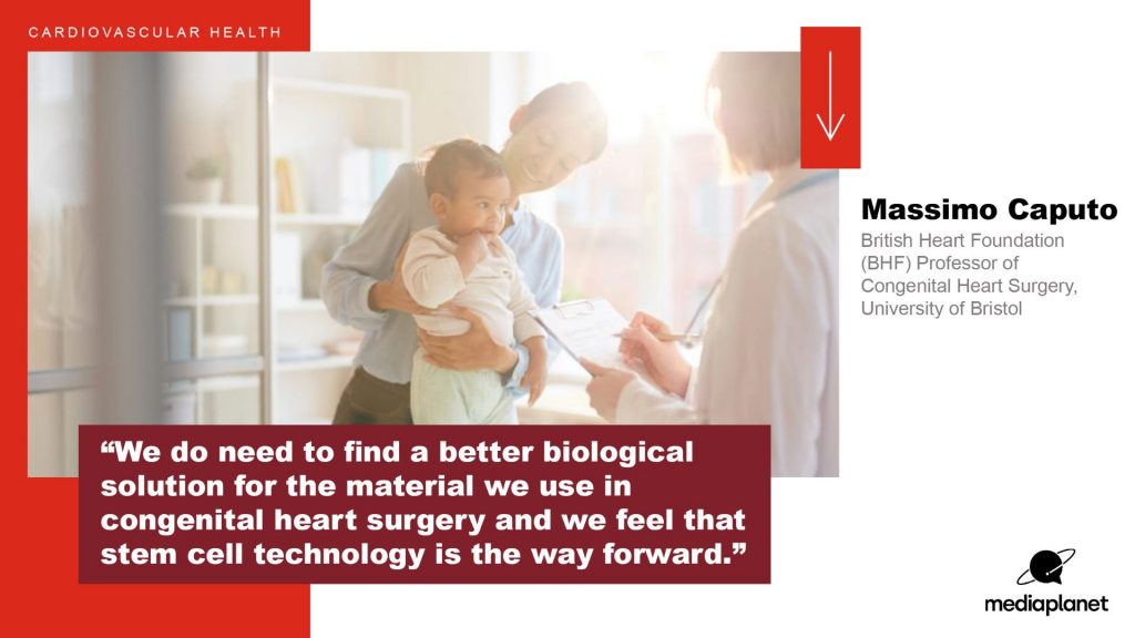
Massimo Caputo, BHF Professor of Congenital Heart Surgery at the Bristol Heart Institute, University of Bristol, said: “For years families have come to us asking why their child needs to have heart surgery time and time again. Although each operation can be lifesaving, the experience can put an unbelievable amount of stress on the child and their parents. We believe that our stem cell patches will be the answer to solve these problems.

“Our ultimate vision in the next decade is to create a paradigm shift in the way doctors treat congenital heart disease, by developing personalised stem cell and genetically-engineered treatments for the most complex of heart defects.”

Dr Sonya Babu-Narayan, Associate Medical Director at the British Heart Foundation, added: “If successful, this new stem cell therapy that acts like a healing plaster could revolutionise the results of heart surgery for children and adults living with congenital heart disease.

“It could offer a solution that means their heart is mended once and forever in a single operation, preventing people from facing a future of repeated surgeries and giving them the gift of a happier and healthier life.”

The work was partly funded by the National Institute of Health and Care (NIHR) Bristol Biomedical Research Centre (NIHR Bristol BRC), a partnership between University Hospitals Bristol and Weston NHS Foundation Trust (UBHW) and the University of Bristol.

New multidisciplinary study could help doctors to predict heart failure

Nearly half of people diagnosed with advanced heart failure (HF) following a major heart attack die within five years. But research supported by GW4 Alliance funding could lead to earlier prediction and intervention – improving life expectancy.

(more…)

TV watching linked with potentially fatal blood clots

Take breaks when binge-watching TV to avoid blood clots, say scientists. The warning comes as a study reports that watching TV for four hours a day or more is associated with a 35 per cent higher risk of blood clots compared with less than 2.5 hours. The University of Bristol research is published today in the European Journal of Preventive Cardiology, a journal of the ESC.

The study examined the association between TV viewing and venous thromboembolism (VTE). VTE includes pulmonary embolism (blood clot in the lungs) and deep vein thrombosis (blood clot in a deep vein, usually the legs, which can travel to the lungs and cause pulmonary embolism).

To conduct the study, the University of Bristol researchers conducted a systematic review to collect the available published evidence on the topic and then combined the results using a process called meta-analysis.

Lead author, Dr Setor Kunutsor from the University’s Bristol Medical School explained:

“Combining multiple studies in a meta-analysis provides a larger sample and makes the results more precise and reliable than the findings of an individual study.”

The analysis included three studies with a total of 131,421 participants aged 40 years and older without pre-existing VTE. The amount of time spent watching TV was assessed by questionnaire and participants were categorised as prolonged viewers (watching TV at least four hours per day) and never/seldom viewers (watching TV less than 2.5 hours per day).

The average duration of follow-up in the three studies ranged from 5.1 to 19.8 years. During this period, 964 participants developed VTE. The researchers analysed the relative risk of developing VTE in prolonged versus never/seldom TV watchers. They found that prolonged viewers were 1.35 times more likely to develop VTE compared to never/seldom viewers.

The association was independent of age, sex, body mass index (BMI) and physical activity. Dr Kunutsor said:

“All three studies adjusted for these factors since they are strongly related to the risk of VTE; for instance, older age, higher BMI and physical inactivity are linked with an increased risk of VTE.

“The findings indicate that regardless of physical activity, your BMI, how old you are and your gender, watching many hours of television is a risky activity with regards to developing blood clots.

“Our study findings also suggested that being physically active does not eliminate the increased risk of blood clots associated with prolonged TV watching,” said lead author Dr Kunutsor. “If you are going to binge on TV you need to take breaks. You can stand and stretch every 30 minutes or use a stationary bike. And avoid combining television with unhealthy snacking.”

Dr Kunutsor noted that the findings are based on observational studies and do not prove that extended TV watching causes blood clots.

Regarding the possible reasons for the observed relationship, he said:

“Prolonged TV viewing involves immobilisation which is a risk factor for VTE. This is why people are encouraged to move around after surgery or during a long-haul flight. In addition, when you sit in a cramped position for long periods, blood pools in your extremities rather than circulating and this can cause blood clots. Finally, binge-watchers tend to eat unhealthy snacks which may lead to obesity and high blood pressure which both raise the likelihood of blood clots.”

Dr Kunutsor concluded:

“Our results suggest that we should limit the time we spend in front of the television. Long periods of TV watching should be interspersed with movement to keep the circulation going. Generally speaking, if you sit a lot in your daily life – for example your work involves sitting for hours at a computer – be sure to get up and move around from time to time.”

Read the paper

Television viewing and venous thrombo-embolism: a systematic review and meta-analysis in the European Journal of Preventive Cardiology by Setor Kunutsor, Richard Dey and Jari Laukkanen.

Further information

Authors’ note:

The findings are based on observational studies, so they do not prove cause and effect.

These findings are based on the results of only three studies. More studies are needed to confirm or refute the findings.

Welcome to the BHI Newsletter Autumn 2021

Professor Andy Judge, Head of Section for Cardiovascular Surgery and Vascular Biology, reflects on recent successes.

With the new academic year underway, we welcome our new cohorts of postgraduate taught master’s degree students for the MSc in Perfusion Science and the MSc in Translational Cardiovascular Medicine (TCM). The teaching sessions are now back in person for the campus based TCM students and Perfusion students, and our student numbers are good.

I’m pleased to report that the labs, which closed for three months at the start of the pandemic, have been open since June 2020. Staff are gradually returning to the office at Level 7 of the BRI as the University has launched its blended working trial policy, and we are now seeing more staff in both the labs and offices.

Within our section for Cardiovascular Surgery and Vascular Biology at the University, we are delighted to have been able to appoint staff to new posts. Dr Kerry Wadey has been appointed as Lecturer in Cardiovascular Medicine on an open-ended core funded post. Francesco Paneni has been appointed as a Professor in Cardiology and Tom Johnson as an Associate Professor in Cardiology and they are expected to begin working with us at the start of 2022.

A number of staff in our section have had success following the annual University promotions procedure. Jason Johnson has been promoted to Professor of Cardiovascular Pathology, and Umberto Benedetto becomes Professor of Cardiac Surgery. Staff from the Teaching and
Learning for Health Professionals (TLHP) programme have recently joined our department of Translational Health Sciences and come under
the umbrella of our cardiovascular section, where we are delighted to congratulate Andrew Blythe on being promoted to Professor of Medical Education in this year’s annual promotion procedure.

Huge congratulations to all those recently promoted: we wish them continued success in their careers.

Bristol Heart Institute researchers feature in World Heart Day campaign


Four Bristol Heart Institute researchers have shared their work in a campaign to mark World Heart Day, 29 September 2021.

The interviews, which appear in the Guardian newspaper’s Cardiovascular Health supplement, cover a broad aspect of the BHI’s work, from tissue engineering to population health.

BHI Director, Professor Gianni Angelini, outlines the BHI’s innovative link between clinicians and scientists, citing the combination of people from different disciplines – basic scientists, clinicians, engineers, epidemiologists and statisticians – as one of the Institute’s key strengths.

Professor of Congenital Cardiac Surgery, Massimo Caputo, talks about how tissue engineering and stem cell therapies could lead to better treatment options for young patients with congenital heart disease.

Dr Giovanni Biglino, Senior Lecturer in Biostatistics, explains how patients and the public are playing an increasing role in cardiovascular research through closer collaborations with clinicians, researchers and artists. This is supporting clinical training and medical research as well as helping patients have a better understanding, and acceptance, of their heart conditions.

Finally, Deborah Lawlor, Professor of Epidemiology, talks about using population health data to identify causes of cardiovascular disease and to predict who is at risk of it. which can lead to prevention, early detection and effective treatment of disease.

See the full BHI World Heart Day campaign

BHI scientist wins national science image contest with heart vessel image

Recreating heart blood vessels
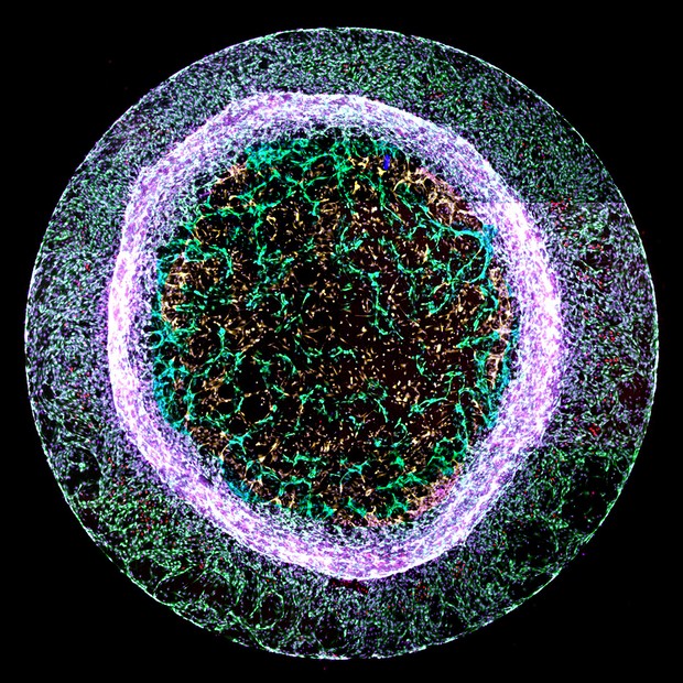
Recreating heart blood vessels. Image credit : Dr Elisa Avolio

A scientist from the Bristol Heart Institute has won the British Heart Foundation’s (BHF) annual ‘Reflections of Research’ image competition. Where science and art collide, the competition challenges BHF-funded scientists to showcase their state-of-the-art heart and circulatory disease research through the generation of captivating images.

Dr Elisa Avolio’s entry ‘recreating heart blood vessels’ was chosen as this year’s judges’ winner. Although at first glance it appears to resemble a luminous jelly fish, the image shows new blood vessel-like structures – pictured in green in the centre – sprouting from a 3D gel.

Dr Avolio of the Bristol Medical School created the structures using a mixture of two types of heart cells – cardiac endothelial cells, which line the inside of every blood vessel, and pericytes, which ‘hug’ the outside of blood vessels to support the vessel and help it function.

During a heart attack, the arteries that supply blood to the heart are blocked, cutting off blood flow. The area of the heart starved of blood and oxygen dies, and it no longer functions to help the heart pump blood around the body. Dr Avolio is researching ways to encourage the formation of new blood vessels to replace those that have died, to restore blood supply to damaged areas of the heart.

Dr Avolio, a post-doctoral research associate, said:

“It is fantastic to have won this year’s Reflections of Research competition. Each year the entries display such variety in the BHF’s work to support heart and circulatory disease research.

“By recreating models of the heart blood vessels, we can see how the cells in blood vessel walls interact with and talk to other cells. This knowledge, along with understanding what molecules promote or block the formation of blood vessels, could be used in the future to develop new treatments for patients after a heart attack.”

Dr Neil Dufton, Lecturer in Inflammatory Sciences at Queen Mary University of London, was this year’s guest judge. He said:

“All of the images shortlisted in this year’s competition offer a stunning glimpse into the cutting-edge work being carried out by BHF scientists.

“The winning image is truly eye-catching. The chaotic mixture of different cells around the outside contrasts perfectly with a ‘through the looking glass’ moment where we see new and exquisitely detailed blood vessels forming in the centre.”

Dr Charmaine Griffiths, Chief Executive at the British Heart Foundation, was also a competition judge. She added:

“All of this year’s entries beautifully capture aspects of the heart and circulatory system, bringing to life the challenges that BHF scientists work tirelessly to solve.

“The images show how far we’ve come over 60 years of BHF research, and would have been barely imaginable to our founders. I love the winning image not just because of its circular beauty, but also because of the hope it represents for the future of healing damaged hearts.”

Early adulthood education and employment experiences play independent role in later life cardiovascular health

New research has found that education and employment experiences in early adulthood contribute to cardiovascular health inequalities in later life, independent of occupation and family income in mid-adulthood. The findings, published today in the Journal of Epidemiology and Community Health, involve researchers from the University of Cambridge, University of Bristol and UCL Social Research Institute.

There are important differences in health between different sectors of our society, with those who are less educated and in lower status jobs shown to be less healthy and have shorter life expectancy on average than the more privileged. While early adulthood is an important time for both the development of adult socioeconomic position and for development of behaviours related to cardiovascular health, until now the degree to which early adulthood socioeconomic trajectories contribute directly to health differences observed in later life has not been clear.

Researchers from the University of Cambridge, University of Bristol and UCL Social Research Institute analysed health and socioeconomic data collected over several decades from over 12,000 members of the 1970 British Birth Cohort, to determine the contribution of early adulthood to differences in cardiovascular health in mid-adulthood. The scientists used a data-driven method to divide the population into different socioeconomic trajectory groups based on their participation in education, different job types, unemployment or economic inactivity across early adulthood (ages 16-24). They studied the association of these groups with cardiovascular risk factors at age 46, including blood pressure, cholesterol levels, waist circumference. To determine if the association of early adulthood socioeconomic trajectories with cardiovascular health was mediated by socioeconomic status later in life, they examined how correcting for occupation or family income at age 46 affected the link.

Professor Kate Tilling from the MRC Integrative Epidemiology Unit at the University of Bristol, and senior author on the paper, said:

“Measuring socioeconomic position in early adulthood has always been difficult as this is a period of transition when most people’s occupations change over time. The method we’ve developed provides a flexible way to identify early adulthood socioeconomic position, and we hope that it will be used in future to answer other research questions related to this period of life.”

The researchers found that those who spent a longer time in education, going on to employment in professional or managerial roles during early adulthood, had better cardiovascular health more than 20 years later (at age 46) than other groups. Importantly this association wasn’t entirely because of a higher income or higher level job at age 46, suggesting an independent and long-term association of early adulthood influences with health.

The findings indicate that that material factors in mid-adulthood do not contribute to the pathway through which early adulthood socioeconomic trajectory affects mid-life health, and the authors suggest that the development of health behaviours or psychosocial factors such as stress, depression, and job control in early adulthood may play an important role.

Dr Eleanor Winpenny from the MRC Epidemiology Unit at the University of Cambridge, and first author on the paper, said:

“We found that an individual’s education and employment experiences in early adulthood had a far larger impact on measures of cardiovascular health more than twenty years later than their occupation or income at that time did.

“These results suggest that we need to provide more support for young adults to allow healthy development into middle age and prevent disease in later life. Given the added disadvantage to young adults as a result the current coronavirus pandemic, there is an urgent need to understand and mitigate the effect these circumstances may be having on their future health.”

The research was funded by the Medical Research Council and the Centre for Diet and Activity Research (CEDAR), a UKCRC Public Health Research Centre of Excellence. Funding from the British Heart Foundation, Cancer Research UK, Economic and Social Research Council, Medical Research Council, the National Institute for Health Research, and Wellcome, under the auspices of the UK Clinical Research Collaboration.

Paper

Early adulthood socioeconomic trajectories contribute to inequalities in adult cardiovascular health, independently of childhood and adulthood socioeconomic position‘ by Winpenny, E. et al in the Journal of Epidemiology and Community Health

The beating heart of Royal Fort Garden

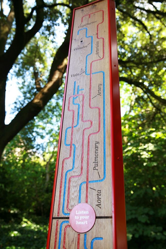
Bristol Heart Institute interpretation sign

The new interpretation sign for the University of Bristol brings a whole new meaning to the phrase ‘living statue’.

The solid oak monolith is the third instalment in a series of iconic totems dedicated to the University’s research institutes. This sign embodies the work of the Bristol Heart Institute and has been designed to reflect the relationship between human life and nature. Joining many other works of art in the Royal Fort Garden this piece stands proud at three metres high, and if you get close enough you can hear the low thud of a heartbeat coming from deep within the structure day and night.

Previous designs highlighted the work of the Bristol Population Health and the Bristol Bio Design Institutes.

The new monolith is built from four solid oak panels which have been laser cut and hand painted to represent a human cardiovascular system. Following a couple of years of pandemic enforced hiatus, it’s great to add to the collection and celebrate the work of the Bristol Heart Institute in such a creative way.  

(Photo by Green Hat. Words adapted from text supplied by Green Hat)

Cardiovascular researchers visit Bristol primary school

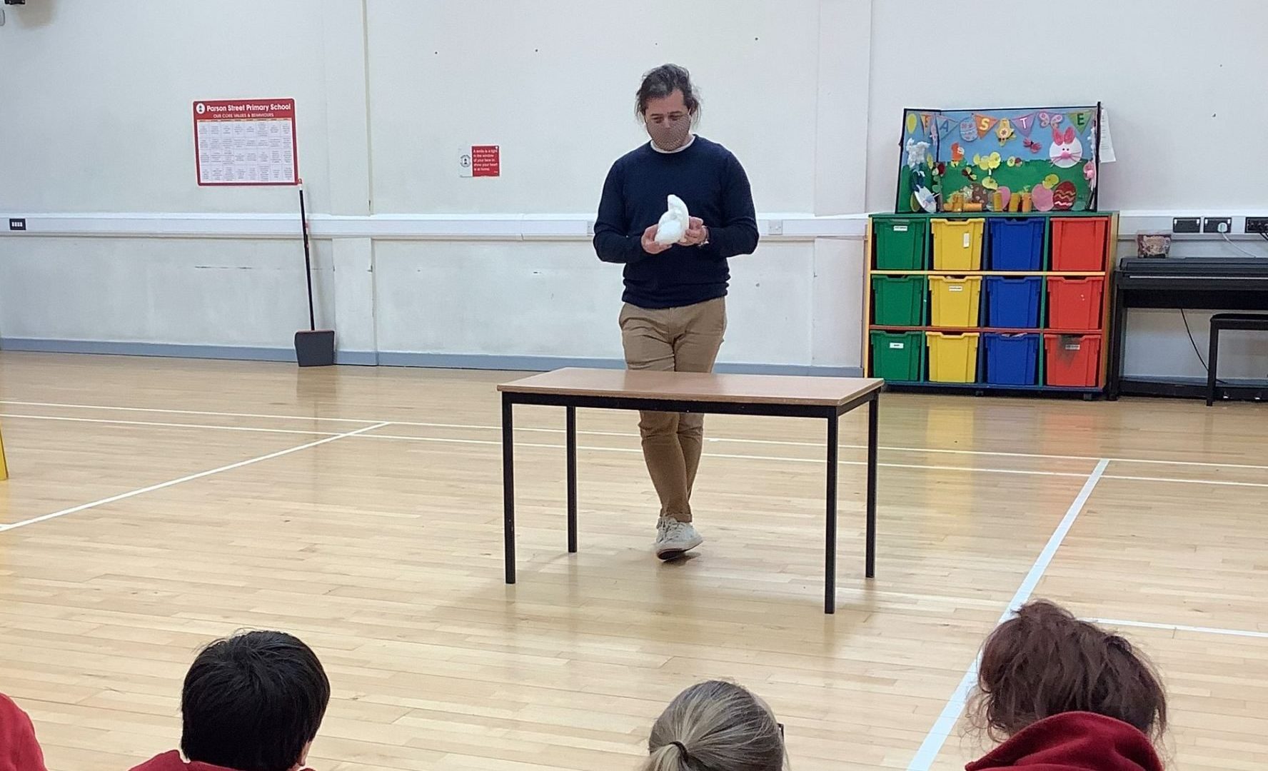
Giovanni Biglino shows Year 6 students a 3D printed heart model

Drs Lucy Culliford, Andrew Shearn and Giovanni Biglino took part in an outreach activity at Parson Street Primary School in Bedminster, south Bristol. in May 2021.

The visit was organised by Lucy on behalf of the Bristol Trials Unit. The group explained some of their research to the Year 6 students (aged 10 and 11) and, as the visit happened in the same week as Clinical Trials Day, it was also an opportunity for Lucy to ask the students to think about clinical studies, the idea of randomisation and ‘what is a clinical trial?’. The group showed the THERMIC 3 animation video, which was developed as part of the TRECA study exploring children’s and young people’s engagement with clinical trials.

Giovanni gave an overview of the technology involved in 3D printing and Andrew brought a series of heart models, both adult and paediatric, including examples of babies’ hearts with congenital heart disease, which sparked some stimulating conversations with the 53 students who attended.

They explained that 3D printing technology can produce models of human organs using scans taken during routine visits to the hospital. Bristol Royal Hospital for Children use this technique to create heart models from patients with congenital heart disease. Being able to hold a life-size model of the patient’s heart can complement information the surgeon or cardiologist can get from medical imaging – for example, they can practice aspects of the surgery or decide the best route to access a specific part of the heart. The models are also used when explaining the details of the case to a patient or a parent.

The children asked lots of questions about ‘hole in the hearts’, as some had had siblings with this condition. They also asked how smoking can affect the heart, and if the researchers could show them a video of a beating heart.

Lucy said:

“This is the first time we’ve done a talk in a school as part of our outreach activities around Clinical Trials Day, and it was a real pleasure to talk to the children. They were very engaged and although they had only started learning about the heart the day before, they already knew lots about heart anatomy, and had more questions than we had time to answer!”

How to get involved in schools outreach

If you would like to run a schools outreach activity, contact your local school.

Schools often have science weeks, or may be doing a topic related to cardiovascular research. For example, Parson Street Primary School use the Cornerstones curriculum and the year 6s had just started the ‘blood heart’ topic – the following week they had a visitor demonstrating a heart dissection.