BHI scientist wins national science image contest with heart vessel image

Recreating heart blood vessels
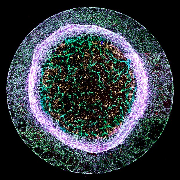
Recreating heart blood vessels. Image credit : Dr Elisa Avolio

A scientist from the Bristol Heart Institute has won the British Heart Foundation’s (BHF) annual ‘Reflections of Research’ image competition. Where science and art collide, the competition challenges BHF-funded scientists to showcase their state-of-the-art heart and circulatory disease research through the generation of captivating images.

Dr Elisa Avolio’s entry ‘recreating heart blood vessels’ was chosen as this year’s judges’ winner. Although at first glance it appears to resemble a luminous jelly fish, the image shows new blood vessel-like structures – pictured in green in the centre – sprouting from a 3D gel.

Dr Avolio of the Bristol Medical School created the structures using a mixture of two types of heart cells – cardiac endothelial cells, which line the inside of every blood vessel, and pericytes, which ‘hug’ the outside of blood vessels to support the vessel and help it function.

During a heart attack, the arteries that supply blood to the heart are blocked, cutting off blood flow. The area of the heart starved of blood and oxygen dies, and it no longer functions to help the heart pump blood around the body. Dr Avolio is researching ways to encourage the formation of new blood vessels to replace those that have died, to restore blood supply to damaged areas of the heart.

Dr Avolio, a post-doctoral research associate, said:

“It is fantastic to have won this year’s Reflections of Research competition. Each year the entries display such variety in the BHF’s work to support heart and circulatory disease research.

“By recreating models of the heart blood vessels, we can see how the cells in blood vessel walls interact with and talk to other cells. This knowledge, along with understanding what molecules promote or block the formation of blood vessels, could be used in the future to develop new treatments for patients after a heart attack.”

Dr Neil Dufton, Lecturer in Inflammatory Sciences at Queen Mary University of London, was this year’s guest judge. He said:

“All of the images shortlisted in this year’s competition offer a stunning glimpse into the cutting-edge work being carried out by BHF scientists.

“The winning image is truly eye-catching. The chaotic mixture of different cells around the outside contrasts perfectly with a ‘through the looking glass’ moment where we see new and exquisitely detailed blood vessels forming in the centre.”

Dr Charmaine Griffiths, Chief Executive at the British Heart Foundation, was also a competition judge. She added:

“All of this year’s entries beautifully capture aspects of the heart and circulatory system, bringing to life the challenges that BHF scientists work tirelessly to solve.

“The images show how far we’ve come over 60 years of BHF research, and would have been barely imaginable to our founders. I love the winning image not just because of its circular beauty, but also because of the hope it represents for the future of healing damaged hearts.”

New research lifts the lid on cardiac microvascular dysfunction

New research has shown abnormalities in the tiny blood vessels of human hearts in regions well beyond the large arteries with atherosclerotic blockages that trigger the need for stents or bypass surgery. The findings could lead to the development of new treatments for patients with angina-like symptoms without blockages or those recovering from a heart attack or unexplained heart failure.

Normal intrinsic constriction of these micro-arteries in response to changing blood pressure is called myogenic (automatic) tone. Myogenic tone controls blood flow distribution within the heart muscle, and in other parts of the human body.

Current heart scans can identify blockages in large coronary arteries, but they are unable to show these tiny, hair size micro-arteries in patients, making it impossible to diagnose poor myogenic tone, which is thought to develop independent of disease in the larger arteries. This study used tissue biopsies to study the function, structure and alterations in pathways in the micro-arteries that link to abnormalities in myogenic tone.

The study, led by Professor Raimondo Ascione (Clinical Lead) at the University of Bristol and Professor Kim Dora (Basic Science Lead) at the University of Oxford, and funded by the BHF, is published in Cardiovascular Research.

The research team took small heart samples, that are otherwise discarded, from 88 patients with no large coronary artery blockages and undergoing valvular cardiac surgery at the Bristol Heart Institute. In addition, cardiac samples were obtained from three human organ donors from the Newcastle Institute of Transplantation Tissue Biobank and 45 pigs treated at the University of Bristol Translational Biomedical Research Centre (TBRC).

The research team found that 44 per cent of the micro-arteries from patients had abnormal myogenic tone despite retaining their cell viability. This abnormality was associated with an excessive presence of a molecule called caldesmon within the muscle cells in the wall of the abnormal micro-arteries and with poor alignment of these contracting cells compared to micro-arteries with normal myogenic tone from the other 66 per cent of patients, and all the organ donors and pigs.

Abnormalities in the micro-arteries affects the blood supply within the beating heart, and other organs in the body, affecting people’s quality of life and life expectancy.

The findings offer new insights on coronary microvascular dysfunction that could predate the development of clinically known heart disease such as heart failure.

Professor Raimondo Ascione, NHS Consultant Cardiac Surgeon and Head of the TBRC at the University of Bristol, said:

“It has been a pleasure to work with Professor Dora on this landmark study over the last seven years. No study had focused on ex vivo poor myogenic tone of the cardiac microcirculation before. These tiny arteries are sited deep within the cardiac wall, well beyond the blocked arteries we treat in the NHS with stents or bypass surgery and cannot be seen with a naked eye.

“Our study lifts the lid on cardiac microvascular dysfunction. It could help to develop new treatments to help patients with angina-like symptoms without coronary blockages, or those recovering from a heart attack or unexplained heart failure.”

Kim Dora, Professor of Microvascular Pharmacology at the University of Oxford, explained:

“I am so excited with the results of this study and the excellent teamwork with Professor Ascione in Bristol.  Not only will our findings enhance the development of new medical treatments and possibly new patient imaging modalities, but they represent a new ex-vivo research model for thousands of scientists globally working on microvascular dysfunction in the heart and other organs.”

Professor Jeremy Pearson, Associate Medical Director at the British Heart Foundation, added:

“This study is the first to develop techniques to understand the links between the structure of micro-arteries and impaired myogenic tone, representing the outcome of years of painstaking work to develop the methods and apply them to micro-arteries from human hearts. The findings provide new information that will help to develop treatments for the many patients whose angina occurs without significant narrowing of their coronary arteries.”

There is now a new area of research that confirms thousands of patients, mostly postmenopausal women, have angina-like symptoms despite their coronary angiogram showing no obvious blockages of the large epicardial arteries in the heart that are usually treated with stent or bypass. Other patients seem to develop heart failure associated with either the contraction or the relaxation of their heart for no obvious reasons.

The human coronary micro-arteries the Bristol and Oxford team has studied in the laboratory represent the microvascular area in human organs (lung, heart, brain and elsewhere) where COVID-19 has caused most of the problems during the ongoing pandemic.

Paper

Human coronary microvascular contractile dysfunction associates with viable 2 synthetic smooth muscle cells‘ by Kim A Dora, Raimondo Ascione et al in Cardiovascular Research [open access]

Early adulthood education and employment experiences play independent role in later life cardiovascular health

New research has found that education and employment experiences in early adulthood contribute to cardiovascular health inequalities in later life, independent of occupation and family income in mid-adulthood. The findings, published today in the Journal of Epidemiology and Community Health, involve researchers from the University of Cambridge, University of Bristol and UCL Social Research Institute.

There are important differences in health between different sectors of our society, with those who are less educated and in lower status jobs shown to be less healthy and have shorter life expectancy on average than the more privileged. While early adulthood is an important time for both the development of adult socioeconomic position and for development of behaviours related to cardiovascular health, until now the degree to which early adulthood socioeconomic trajectories contribute directly to health differences observed in later life has not been clear.

Researchers from the University of Cambridge, University of Bristol and UCL Social Research Institute analysed health and socioeconomic data collected over several decades from over 12,000 members of the 1970 British Birth Cohort, to determine the contribution of early adulthood to differences in cardiovascular health in mid-adulthood. The scientists used a data-driven method to divide the population into different socioeconomic trajectory groups based on their participation in education, different job types, unemployment or economic inactivity across early adulthood (ages 16-24). They studied the association of these groups with cardiovascular risk factors at age 46, including blood pressure, cholesterol levels, waist circumference. To determine if the association of early adulthood socioeconomic trajectories with cardiovascular health was mediated by socioeconomic status later in life, they examined how correcting for occupation or family income at age 46 affected the link.

Professor Kate Tilling from the MRC Integrative Epidemiology Unit at the University of Bristol, and senior author on the paper, said:

“Measuring socioeconomic position in early adulthood has always been difficult as this is a period of transition when most people’s occupations change over time. The method we’ve developed provides a flexible way to identify early adulthood socioeconomic position, and we hope that it will be used in future to answer other research questions related to this period of life.”

The researchers found that those who spent a longer time in education, going on to employment in professional or managerial roles during early adulthood, had better cardiovascular health more than 20 years later (at age 46) than other groups. Importantly this association wasn’t entirely because of a higher income or higher level job at age 46, suggesting an independent and long-term association of early adulthood influences with health.

The findings indicate that that material factors in mid-adulthood do not contribute to the pathway through which early adulthood socioeconomic trajectory affects mid-life health, and the authors suggest that the development of health behaviours or psychosocial factors such as stress, depression, and job control in early adulthood may play an important role.

Dr Eleanor Winpenny from the MRC Epidemiology Unit at the University of Cambridge, and first author on the paper, said:

“We found that an individual’s education and employment experiences in early adulthood had a far larger impact on measures of cardiovascular health more than twenty years later than their occupation or income at that time did.

“These results suggest that we need to provide more support for young adults to allow healthy development into middle age and prevent disease in later life. Given the added disadvantage to young adults as a result the current coronavirus pandemic, there is an urgent need to understand and mitigate the effect these circumstances may be having on their future health.”

The research was funded by the Medical Research Council and the Centre for Diet and Activity Research (CEDAR), a UKCRC Public Health Research Centre of Excellence. Funding from the British Heart Foundation, Cancer Research UK, Economic and Social Research Council, Medical Research Council, the National Institute for Health Research, and Wellcome, under the auspices of the UK Clinical Research Collaboration.

Paper

Early adulthood socioeconomic trajectories contribute to inequalities in adult cardiovascular health, independently of childhood and adulthood socioeconomic position‘ by Winpenny, E. et al in the Journal of Epidemiology and Community Health

The beating heart of Royal Fort Garden

Bristol Heart Institute interpretation sign

The new interpretation sign for the University of Bristol brings a whole new meaning to the phrase ‘living statue’.

The solid oak monolith is the third instalment in a series of iconic totems dedicated to the University’s research institutes. This sign embodies the work of the Bristol Heart Institute and has been designed to reflect the relationship between human life and nature. Joining many other works of art in the Royal Fort Garden this piece stands proud at three metres high, and if you get close enough you can hear the low thud of a heartbeat coming from deep within the structure day and night.

Previous designs highlighted the work of the Bristol Population Health and the Bristol Bio Design Institutes.

The new monolith is built from four solid oak panels which have been laser cut and hand painted to represent a human cardiovascular system. Following a couple of years of pandemic enforced hiatus, it’s great to add to the collection and celebrate the work of the Bristol Heart Institute in such a creative way.  

(Photo by Green Hat. Words adapted from text supplied by Green Hat)