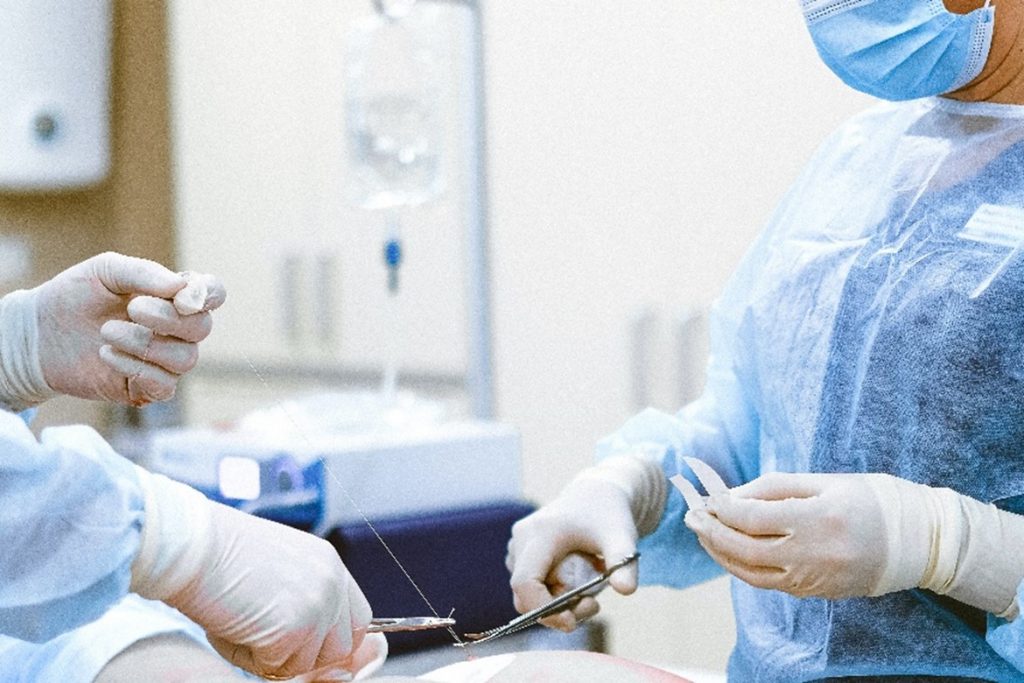
Cardiac surgeons could in the future be conducting procedures virtually before even stepping into an operating theatre. A research team from UWE Bristol’s Big Data lab and Faculty of Health and Applied Sciences (HAS) is developing technology that uses artificial intelligence (AI), augmented reality (AR) and virtual reality (VR) to assist cardiac surgeons in planning and preparing for complex keyhole heart valve surgery.
The team is initially collaborating with the Bristol Heart Institute (BHI), a Specialist Research Institute at the University of Bristol, whose surgeons will test the system when preparing for minimally invasive cardiac valve surgery (MICVS).
Compared to conventional open-heart surgery involving cutting through the breastbone to reach the heart, MICVS is less intrusive as the heart is accessed through smaller incisions using endoscopic instruments. And patient recovery time is generally quicker after this keyhole surgery.
However, MICVS is complex and requires hours of pre-operative planning and preparation.
Dr Hunaid Vohra, Consultant Cardiac Surgeon and Honorary Senior Lecturer and Researcher at the BHI, who is collaborating with UWE Bristol, said:
“In the operating room, despite pre-planning, it is currently very common to find unexpected challenges, as every patient’s height, weight and heart-lung anatomy is different. And patients’ frailty varies.
“Mixed Reality and AI will enhance our ability to prevent the conversion of a keyhole heart valve operation to an open heart surgery, avoiding two sets of scars, and delay in recovery.”
Surgeons will initially be able to use the system’s AI to tap into the patient’s medical data to predict the risks associated with the procedure. The likelihood of adverse events is then presented to the surgeon on a HoloLens using AR.
Next, the surgeon will have access to AR technology to show a patient a 3D version of their heart and explain the procedure to them via headsets.
Dr Muhammad Bilal, Associate Professor of Big Data and Artificial Intelligence at UWE Bristol and leading the research team, said:
“Most terms surgeons use to describe heart surgery during consultation draw a blank from patients and this system makes the explanation task much clearer and easier.”
Incorporated in the system is also a pre-operative logistics element that optimises operation planning. This will assist medical teams in preparing the right instruments and materials, and booking the appropriate operating theatre and hospital beds, among other tasks.
Crucially, the software’s virtual planning feature will provide surgeons with access to a complete digital version of the patient, enabling them to perform the entire operation beforehand on a replica of the patient’s thoracic cavity. This will include ‘what-if’ scenarios to determine the most optimal and personalised surgical strategies.
Finally, in collaboration with UWE Bristol’s Centre for Print Research, surgeons performing very complex cases will be allowed to order a bespoke 3D printed model of the patient’s thoracic cavity mimicking organs, veins, and blood flow to simulate the procedure on a synthetic body.
Dr Vohra said:
“This will enable us to practise before the actual operation and minimise the potential for things to go wrong on the day. Overall, we are excited to be involved in this technology, which could spell the future for highly successful minimally invasive procedures of this type in adults and babies.”
Dr Bilal added:
“Currently, the practice of MICVS is limited to a small group of surgeons in the world. This technology-enabled guidance promises to increase the number of doctors able to perform these operations, providing wider access to the general population.
“There are significant engineering challenges to be resolved before this technology can be rolled out into the NHS but our collaboration with the BHI provides a perfect testing ground.”

